Tuesday, November 23, 2010

Human blood progenitors made from skin

Following earlier studies showing that mouse fibroblasts could be converted into neurons and muscle cells without the need to create first iPS, a recent paper in Nature presented the first direct conversion of human skin cells into blood subtypes.
Szabo et al. (7 Nov 2010; doi:10.1038/nature09591) transduced human skin fibroblasts with the pluripotency factor OCT4 and grew them with different cytokines to convert them into hematopoietic progenitors and mature blood cells. After a few days, the cells expressed the pan- haematopoietic factor CD45 and eventually gave rise to all blood cell types (myeloid, erythroid and megakaryocytic lineages), depending on the cytokines added to the medium.

The hemotopoietic differentiation medium used in this study to derive blood progenitors was made with cytokines from R&D Systems:

Friday, November 19, 2010

Embryonic stem cell defined media: culturing goes from art to science

A team of researchers at the University of Wisconsin-Madison reports the development of a fully defined embryonic or pluripotent stem cells culture system.
[R&D Systems has an excellent defined media, StemXVivo culture matrix, composed of a mixture of recombinant human adhesion molecules for the culture of stem/progenitor cells.]
Writing this week (Nov. 14, 2010) in Nature Methods, a team led by Laura Kiessling, unveiled an inexpensive system that takes much of the guess work out of culturing the all-purpose cells.
"It's a technology that anyone can use," says Kiessling. "It's very simple."
The new culture system utilizes a synthetic, chemically made substrate of protein fragments, peptides, which have an affinity for binding with stem cells. Used in combination with a defined growth medium, the system devised by the Wisconsin team can culture cells in their undifferentiated states for up to three months and possibly longer. The system, according to the new report, also works for induced pluripotent stem cells, the adult cells genetically reprogrammed to behave like embryonic stem cells.


Figure 3
(Lim et al.)
: Synthetic surfaces support the long-term culture of pluripotent stem cells.

(a) Immunostaining of H9 hES cells cultured for three months in mTeSR with ROCK inhibitor on the synthetic surface for Oct-4 and SSEA-4, and stained with DAPI. Scale bar, 200 μm. (b,c) Immunostaining of DF19-9 7T iPS cells cultured for 2.5 months in mTeSR with ROCK inhibitor on the synthetic surface for nanog and E-cadherin (b) or for Sox2 and Tra-1-60


R&D Systems reagents used in this study:

recombinant proteins:
Activin A
BMP-4
Noggin

Plates:
Vitronectin-coated plates

Antibodies:
Oct-4
Nanog
Sox-2
β-III tubulin
FoxA2
Fatty acid binding protein 4
Alkaline phosphatase, APC-conjugated
SSEA-1, PE–conjugated


Joseph R Klim, Lingyin Li, Paul J Wrighton, Marian S Piekarczyk & Laura L Kiessling. A defined glycosaminoglycan-binding substratum for human pluripotent stem cells. Nature Methods, 14 November 2010 DOI: 10.1038/nmeth.1532

[News adapted from this UW-Madison press release]

Tuesday, October 19, 2010

Detection of Epithelial-To-Mesenchymal Transition

R&D Systems now offers a Human EMT 3-Color Immunocytochemistry Kit (Catalog # SC026). The kit contains three fluorochrome-conjugated antibodies that can be used together for single-step immunocytochemical assessment of epithelial-to-mesenchymal transition (EMT). EMT is a mechanism by which cells shed their epithelial characteristics and acquire more migratory mesenchymal cell-like properties. This process is essential for embryonic development and wound healing, but is also involved in cancer metastasis. The antibodies included in the kit are NorthernLights 637-conjugated anti-human E-Cadherin, NorthernLights™ 493-conjugated anti-human Vimentin, and NorthernLights 557-conjugated anti-human Snail. Snail is a transcription factor that acts as a master regulator of EMT, while E-Cadherin and Vimentin serve as epithelial and mesenchymal markers, respectively.


Detection of EMT in TGF-beta 1-treated Human Lung Carcinoma Cells using the Human EMT 3-Color Immunocytochemistry Kit. A549 human lung carcinoma cells were either untreated (A) or treated with Recombinant Human TGF-beta 1 (Catalog # 240-B) for 48 hours (B ). The cells were analyzed for an epithelial-to-mesenchymal transition (EMT) by simultaneously staining with antibodies contained in the Human EMT 3-Color Immunocytochemistry Kit (Catalog # SC026 ), including NorthernLights 637-conjugated anti-human E-Cadherin (pseudo-colored gray), NorthernLights 493-conjugated anti-human Vimentin (green), and NorthernLights 557-conjugated anti-human Snail (red). Induction of EMT following TGF-beta 1 treatment was revealed by down-regulation of E-Cadherin and concurrent up-regulation of Vimentin and Snail.

For more information on products for EMT research, please visit our website at www.RnDSystems.com/go/EMT

Monday, September 27, 2010

A Mesenchymal-to-Epithelial Transition Initiates and Is Required for the Nuclear Reprogramming of Mouse Fibroblasts

Li et al. Cell Stem Cell, Volume 7, Issue 1, 51-63, 17 June 2010
10.1016/j.stem.2010.04.014

Highlights:
  • Mouse fibroblast reprogramming involves a mesenchymal-to-epithelial transition
  • Sox2, Oct4, and c-Myc suppress TGF-β signaling
  • Klf4 activates multiple epithelial genes including E-cadherin
  • E-cadherin knockdown impairs reprogramming


R&D Systems used in these studies:
R&D

Klf4 ChIP from Figure 4 of Li et al.

Tuesday, August 17, 2010

Tuesday, June 15, 2010

New: APC-conjugated Mouse CCR2 Monoclonal Antibody


Detection of CCR2 by Flow Cytometry. A. L1.2 mouse pro-B cells transfected with mouse CCR2 were stained with APC-conjugated Mouse CCR2 Monoclonal Antibody (Catalog # FAB5538A; filled histogram) or APC-conjugated Rat IgG2B Isotype Control (Catalog # IC013A; open histogram). B. Mouse peripheral blood cells were stained with APC-conjugated Mouse CCR2 Monoclonal Antibody (Catalog # FAB5538A) and PE-conjugated Mouse Integrin alpha M/CD11b Monoclonal Antibody (Catalog # FAB1124P).


R&D Systems now offers an APC-conjugated Mouse CCR2 Monoclonal Antibody (Catalog # FAB5538A) for flow cytometry. CCR2 is a G protein-linked chemokine receptor that preferentially binds monocyte chemo-attractant protein-1 and -3 (MCP-1/CCL2 and MCP-3/CCL7). Cells that respond to the action of MCP-1, and are therefore likely to express CCR2 receptors, include monocytes, T cells, natural killer cells, basophils, mast cells, and dendritic cells. CCR2, along with other chemokine receptors, is involved in the recruitment of leukocytes to sites of inflammation, making it a potential target for inflammatory disorders. In addition, CCR2 can serve as an HIV fusion co-factor, suggesting that it may also serve as a target for inhibiting the HIV infection process. For more information, please visit our website at www.RnDSystems.com or call 1-800-343-7475.

Monday, March 1, 2010

R&D Systems FoxP3 Antibodies and Kits

FoxP3 is a major marker for regulatory T cells (T Regs) and it is a member of the forkhead family of DNA-binding proteins. T Regs are essential regulators of the immune system and important players in autoimmune disorders, transplantation therapy, and cancer. R&D Systems has developed a new FoxP3 polyclonal antibody that is highly specific (Figure 1). This new FoxP3 antibody has been validated for Western blotting (Figure 1), immunohistochemistry (Figure 2), multi-color flow cytometry (Figure 3) and chromatin immunoprecipitation (Figure 4).


check out all R&D Systems FoxP3-related products here

Tuesday, December 8, 2009

New Mouse Natural Killer Isolation kit

We have developed two new kits for the isolation of untouched Natural Killer cells from both mouse and human preparations that achieves levels of purity as high as 95%. Undesired populations are negatively depleted using a cocktail of monoclonal antibodies that specifically react with non-NK cell populations. These cell types are then tagged with magnetic beads and separated from the desired NK cells by magnetic force. A typical isolation is achieved in 45 minutes.

Introducing the new mouse NK Cell Isolation Kit [Catalog# MAGM210]:


Example of enrichment of NK cells from mouse Balb/C splenocytes using MagCellect Mouse NK cell enrichment kit (R&D Systems MAGM210). Cells before (panel A) and after (panel B) enrichment were double-stained with APC-conjugated anti mouse CD49b (DX5) and PE-conjugated anti mouse NKp46 (R&D Systems Cat.# FAB2225P) to specifically label the NK cell population. Note that two small populations with CD49dim/NKp46- and NKp46dim/CD49b- staining are also enriched (panel C)


Phenotypic characterization of MagCellect-isolated mouse NK cells
MagCellect (MAGM210)-isolated NK cells were characterized by NK-specific staining with PE-conjugated anti-mouse NKp46 (R&D Systems Cat.#) and anti-mouse NKG2D (R&D Systems Cat.#), and with APC-conjugated anti-mouse CD49b (R&D Systems Cat.#) antibodies. Cells were analyzed by flow cytometry.

Comparing the new MagCellect NK Isolation kit with other commercially available systems


First, we tested our new MagCellect NK cell kits by comparison to other systems in the market, with typically better or similar results. But while other commercially available kits require a costly set of magnets and supplies (i.e, expensive columns), MagCellect is simpler and more cost-effective

here's how our kit's performance compares to that a column-based magnetic isolation system:
Balb/C mouse NK cells were isolated from 50 million splenocytes using either R&D Systems (MAGM210) or a competitor's column-based magnetic system. All reagents used (kit reagents, magnets, magnetic beads and columns)were from the respective makers. All isolations were performed according to the given manufacturer's instructions.

MagCellect performed similarly or better in all tests, it is faster, and it is more cost-efficient (you don't have to buy expensive disposable columns).


Second, we tested our new MagCellect NK cell kit in combination with other suppliers' magnets.
We realize that some people already invested large amounts of money in costly magnets and supplies from our competitors, so we developed our MagCellect kits to be able to work with optimal performance using different type of magnets and supplies.
Here's how our MagCellect mouse NK Isolation kit (MAGM210) performed using two magnets from other makers, Miltenyi and StemCell Technologies:
Balb/C mouse NK cells were isolated from 50 million splenocytes using R&D Systems NK isolation kit (Cat# MAGM210) used in combination with either A) R&D Systems magnet (Cat# MAG997), B) Miltenyi Biotec magnet (Cat# 130-042-302) and LS columns (Cat# 130-042-401) or C) StemCell Technologies magnet (Cat# 18000)

MagCellect kit was used with excellent results using any of the leading magnets currently in the market. If your lab already owns a Miltenyi Biotec or StemCell Technology magnet, you can safely switch to using MagCellect with optimal results and in a more cost-efficient manner. Unlike other commercially available kits that require a costly set of magnets and supplies, our kits were developed to work with different kind of commercially available magnets and systems, providing more flexibility, simplicity and cost-efficacy.

New Human Natural Killer Cell purification kit

We have developed two new kits for the isolation of untouched Natural Killer cells from both mouse and human preparations that achieves levels of purity as high as 95%. Undesired populations are negatively depleted using a cocktail of monoclonal antibodies that specifically react with non-NK cell populations. These cell types are then tagged with magnetic beads and separated from the desired NK cells by magnetic force. A typical isolation is achieved in 45 minutes.

Introducing the new Human NK Cell Isolation Kit [Catalog# MAGH109]:

A

B

Two examples of enrichment of NK cells from human PBMC using MagCellect NK
cell enrichment kit (R&D Systems MAGH109). Cells before and after enrichment
were double-stained with APC-conjugated anti human CD3 (R&D Systems FAB100A) and PE-conjugated anti human CD56 (R&D Systems FAB2408) to specifically label the NK cell populations. Panel A, Isolation of NK cells from a PBMC prep consisting of mostly CD56dim cells. Panel B, enrichment of both CD56dim and CD56bright from a different PBMC preparation; CD56dim NK cells are shown in green and CD56bright NK cells are shown in blue.


Phenotypic characterization of MagCellect-isolated NK cells

We have used the most recent and established markers and techniques to extensively characterize the isolated NK cells. We show here that the highly pure NK cell populations (both mouse and human) express NK cell-specific markers (i.e., NKp46, Nkp80, NKp30, CD56, NKG2D, KIR3DL1 and NTB-A)


List of R&D Systems APC- or PE-conjugated antibodies used in the phenotypic characterization of isolated NK cells:

Marker- Catalog # (click on the cat# to see more info)
NKp46 - FAB1850A
NKp44 - FAB22491A
NKp80 - FAB1900A
NKp30 - FAB1849P
CD56 - FAB2408A
NTB-A - FAB19081A
NKG2D - FAB139A
NKG2A - FAB1059A
NKG2C - FAB138A
Functionality of MagCellect-isolated NK cells

1) Isolated human NK cells were probed in a degranulation assay by measuring expression of CD107a (LAMP-1) in a flow cytometric assay after stimulation of isolated NK cells with myelogenous leukaemia K562 cells.


Enriched human NK cells were functional as demonstrated with a CD107adegranulation potential assay. NK cells were incubated with HLA class I- K562 target cells at an Effector/Target (E/T) ratio of 2:1 for 3 hours (Ravet, S. et al (2007) Blood 109:4296). Cells were then stained with APC-labeled anti-human CD107a/LAMP1 (R&D Systems Catalog #IC4800A) and analyzed by flow cytometry.

2) Interferon gamma induction on isolated NK cells was assayed


Immobilized Ms x hNKp30 (R&D Systems Inc. catalog #MAB1849) induces IFNγ secretion in IL-2 activated human NK cells. Isotype control did not induce IFNγ secretion. Human peripheral blood NK cells were isolated using MagCellect Human NK cell isolation kit (R&D Systems Inc. catalog #MAGH109). The amount of IFNγ secretion was measured using the human IFNγ kitQuantikine (R&D Systems Inc. catalog #DIF50).


Granzyme expression in intact isolated NK cells


MagCellect (MAGH109)-Isolated NK cells were stained with anti-human NKp46 (Catalog # AF1850) followed by NL493-coupled anti-goat IgG (Catalog # NL003; green), and with anti-human Granzyme B (Catalog # MAB2906) followed by NL557-coupled anti-mouse IgG (Catalog # NL007; red) in the dark. Nuclei were stained with Dapi (blue).
We tested the efficacy of our new MagCellect NK cell kits by comparison to other systems in the market, with typically better or similar results. But unlike other commercially available kits that require a costly set of magnets and supplies (expensive magnets and columns), our kits were developed to work with different kind of commercially available magnets and systems, providing more flexibility, simplicity and cost-efficacy.
We realize that some people already invested large amounts of money in costly magnets and supplies from our competitors, so we developed our MagCellect kits to be able to work with optimal performance using different type of magnets and supplies.

To see how our kits compare to other commercially available isolations systems, and how you can effectively use MagCellect NK Isolation kits with other suppliers magnets and/or column, check out the data presented here.

Monday, October 5, 2009

Hematopoietic stem cells and leukemia stem cells

Video of Irving Weissman from Stanford University School of Medicine on hematopoietic stem cells and leukemia stem cells. Presents some of the findings on CD47 expression and leukemia stem cells we discussed in a previous post.


[This and other high quality presentations can be found on the American Asociation for Cancer Research (AACR) webcasts page.]

Friday, October 2, 2009

Generation of Induced Pluripotent Stem Cells from Human Cord Blood

In the October 2 issue of Cancer Stem Cells two independent groups demonstrate that human cord blood can be reprogrammed to pluripotency. Belmonte and colleagues reprogrammed CD133+ cord blood cells with as few as two transcription factors, Oct4 and Sox2, to generate teratoma-forming human iPSCs. This population reprogrammed with a higher efficiency than fibroblasts. In the second report, Haase, Martin et al. also generate iPSCs from cord blood, and are able to later differentaite the newly reprogrammed cells into functional cardiomyocytes (check out this movie showing their cardiomyocytes beating)


Generation of Induced Pluripotent Stem Cells from Human Cord Blood
Haase, Martin et al.
Cell Stem Cell, Vol. 5, Issue 4, 434-441, 2 October 2009

Generation of Induced Pluripotent Stem Cells from Human Cord Blood Using OCT4 and SOX2
Giorgetti, Belmonte et al.
Cell Stem Cell, Vol. 5, Issue 4, 353-357, 2 October 2009


R&D Systems used in these studies:
R&D


Tuesday, September 29, 2009

Cytoplasmic location of pluripotent stem cell markers and expression of CD44 isoforms in putative breast cancer stem cells


Since expression of pluripotent stem cell markers in tumor-derived cancer stem
cells is controversial, we examined the expression of Oct-3/4, Nanog and SOX2 by immunocytochemistry in tumor spheres derived from the breast cancer cell line MCF7.

In a separate experiment, we analyzed the expression of different CD44 variants typically expressed in cancer cells by flow cytometry. While breast cancer stem cells are typically characterized by their CD44+/CD24low expression, little is known about the expression profiles of CD44 variants in those cells.

Download the poster in high resolution pdf format

(presented at the 2009 American Association for Cancer Research)

Tuesday, September 15, 2009

Yamanaka, Gurdon take the 2009 Lasker Award

John Gurdon of Cambridge University and Shinya Yamanaka of Kyoto University received the 2009 Albert Lasker Basic Medical Research Award for discoveries concerning nuclear reprogramming, the process that instructs specialized adult cells to form early stem cells — creating the potential to become any type of mature cell for experimental or therapeutic purposes.

Gurdon, who at 76 has much better hair than Yamanaka (47), will share with him a prize that is usually seen as a lead up to a Nobel Prize (In the 63-year history of the Lasker prizes, 76 winners also have won a Nobel).


[Read the Lasker Foundation official award justification here]

Gurdon was the first to overturn conventional wisdom by showing that cell development isn’t always a one-way process in which cells go from embryo cells, able to become any other cell type, to unchangeable cells like muscle or nerve cells.
“The prevailing thought was that as cell differentiate, they lose their ability to generate other cells of any kind,” Gurdon told Bloomberg. Gurdon took cells from an adult frog’s gut, inserted the nucleus into an egg cell whose own nucleus had been removed, and created a tadpole with the genetic characteristics of the original frog. His work showed that the adult cells retained the ability to become other cells and that the egg had the power to reawaken those properties.
More than four decades later, Shinya Yamanaka showed that the same results could be achieved without the use of eggs. In 2006, Yamanaka introduced four genes — Oct3/4, Sox2, c-Myc, and Kif4 — into mouse skin cells to transform them into induced pluripotent stem cells —he called them “iPS” cells— which were almost indistinguishable from embryonic stem cells.
“We did it by transferring the nucleus of a cell,” Gurdon said. “Amazingly, he does it by adding genes to the cells and some of them go back to being embryo cells.” "I did not expect it would be possible to do what Yamanaka did.”

The next year Yamanaka published the seminal human iPS paper using the same genes to achieve pluripotency in fibroblast cells:


R&D Systems reagents used in this study:

Tuesday, August 25, 2009

Natural killer cells: Stop, look, listen

On how NK cells balance activating and inhibitory ligands in target cells to decide when to attack.

When a migratory natural killer (NK) cell encounters a potential target cell, it must stop and integrate signals from various sensory inputs to decide whether to proceed with a cytolytic response. Daniel Davis and colleagues show how the formation of a lytic synapse between an NK cell and a target cell, resulting in the polarized secretion of cytolytic granules, is continuously regulated by the balance between signalling through activating and inhibitory receptors.
[from Nature Reviews Immunology 9, 606, September 2009]

Comments from this interesting article:
Natural Killer Cell Signal Integration Balances Synapse Symmetry and Migration. Culley FJ, Johnson M, Evans JH, Kumar S, Crilly R, et al. 2009 PLoS Biol 7(7): e1000159. doi:10.1371/journal.pbio.1000159


R&D  R&D Systems reagents used in that study:

Monday, August 17, 2009

Cancer stem cells news

Cancer Stem Cells (or 'tumor-initiating cells') are in the news again, this time with promising research on potential therapeutics (see NYTimes article here)

A team of scientists including Eric Lander, Robert Weinberg and Charlotte Kuperwasser identified selective inhibitors of Cancer Stem Cells by high-throughput screening (Gupta et al., Cell, August 13 2009) . 32 of the 16,000 chemicals screened selectively went after cancer stem cells.

Scientists from OncoMed Pharmaceuticals, a company dedicated to develop cancer therapies by targeting tumor-initiating cells, published an article in Cell Stem Cell demonstrating anti-cancer activity of a DLL4 blocking antibody in colon and breast cancer models (1). The antibody, the company’s first product candidate, OMP-21M18, is currently in Phase 1 clinical testing.

As we mentioned earlier in this post, blocking monoclonal antibodies directed against CD47 preferentially enabled phagocytosis of acute myeloid leukemia stem cells and inhibited their engraftment in vivo, making CD47 a potential therapeutic target for AML.
The same team led by Irv Weissman has now identified the first human bladder cancer stem cell, and has shown CD47 here again as a key player in allowing cancer cells to escape the body’s natural defenses (Chan et al., PNAS, June 28 2009).

Richard Lock and colleagues have also shown evidence of the elimination of human acute myeloid leukemic stem cells, this time by monoclonal antibody-mediated targeting of CD123, the IL-3 Receptor alpha Chain (Jin et al., Cell Stem Cell (5) 1, July 2009) .

(1) Recombinant human DLL4 from R&D Systems was used in the DLL4 study


R&DDLL4 products from R&D Systems

CD47 products from R&D Systems

IL-3R alpha (CD123) products from R&D Systems

Thursday, August 13, 2009

CD47 expression in Leukemia Stem Cells

Expression of the cell-surface protein CD47 allows some normal cells to avoid phagocytosis by macrophages. Two articles in the July 23 issue of Cell from Irving Weissman's Lab at Stanford University show that elevated CD47 expression by leukemic stem cells inhibits macrophage activity and is an indicator of poor prognosis for patients with acute myeloid leukemia.

CD47 Is Upregulated on Circulating Hematopoietic Stem Cells and Leukemia Cells to Avoid Phagocytosis, Jaiswal et al. Cell 138 (2), 271-285 (2009)

CD47 Is an Adverse Prognostic Factor and Therapeutic Antibody Target on Human Acute Myeloid Leukemia Stem Cells, Majeti et al. Cell, 138 (2), 286-299 (2009)


Click here for a list of CD47 products from R&D Systems
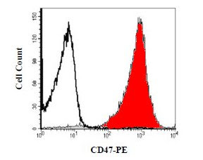
hCD47 histogramHuman lymphocytes were stained with PE-conjugated anti-human CD47 (Catalog # FAB4670P, red histogram) or isotype control (Catalog #IC002P, open histogram).

Wednesday, August 12, 2009

Development and Characterization of a Novel Monoclonal Antibody to Human IL-17A


Download the poster in hi-res pdf format

Although IL-17A is made by CD4+ Th17 cells and there is an ever-increasing body of literature on these cells, other cells make IL-17A, including CD8+ (Tc17) and g-d T cells, NK cells, and neutrophils. There are many factors that contribute to published findings of the percentage of cells producing IL-17A, including the activation or differentiation conditions, time course of activation, and antibody used for detection. It has been published that high levels of IL-17A are secreted by human PBMCs after anti-CD3/CD28-treatment alone for only 24 hrs. We have developed a new monoclonal antibody (Clone 41802) that is specific for human IL-17A. The specificity of this Ab has been rigorously tested. Clone 41802 detects an increase in IL-17A in PMA/ionomycin-treated human PBMCs over resting cells by intracellular flow cytometry. This finding correlated with Western blot data in Th17-differentiated vs resting CD4+ PBMCs. This clone also detects IL-17A in a population of activated CD3+ PBMCs that is also positive for IL-22 and IL-23 R by flow cytometry. Importantly, clone 41802 detects IL-17A in transfectant cells overexpressing human IL-17A, but not in cells overexpressing human IL-17F. In conclusion, clone 41802 is specific for human IL-17A. Although a larger percentage of IL-17A+ cells are detected in activated PBMCs than with other commercially available clones, this indicates an interesting biological finding, not a lack of antibody specificity.